PGx01
Ultra-weit abstimmbarer Pikosekunden-OPA mit hoher Pulsenergie
Diese OPAs sind eine ausgezeichnete Wahl für Forscher, die eine abstimmbare kohärente Lichtquelle mit Pikosekundenpulsen mit hoher Pulsenergie von 193nm im UV bis 16 µm im mittleren IR benötigen.

Entwickelt und hergestellt von:
Features
- Ultraweiter Spektralbereich von 193 bis 16.000 nm
- Hohe Spitzenleistung (> 50 MW), ideal für nichtlineare Spektroskopie
- Schmale Linienbreite < 6 cm⁻¹ (für UV < 9 cm‑1)
- Motorisierte, softwaregesteuertes Wellenlängen-Tuning im Bereich von 193–2.300 nm oder 2.300–16.000 nm
Anwendungen
- Nichtlineare Spektroskopie: Schwingungs-SFG, Oberflächen-SH, Z-Scan
- Pump-Probe-Experimente
- Laserinduzierte Fluoreszenz (LIF)
Anwendungen
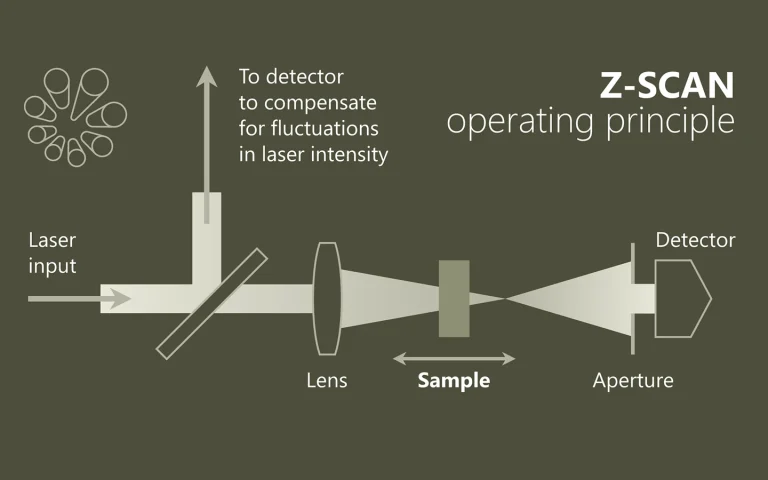
Im Brennpunkt der Nichtlinearität
Im Bereich der Nichtlinearen Optik werden mittels Z-Scan beispielsweise der n2-Index (Kerr-Nichtlinearität) oder der Nichtlineare Absorptionskoeffitzient Δα ermittelt. Über die „Open“ und „Closed“ Methode werden dazu sowohl der Realteil als auch der Imaginär-Teil des Nichtlinearen Brechungs-Indexes gemessen.
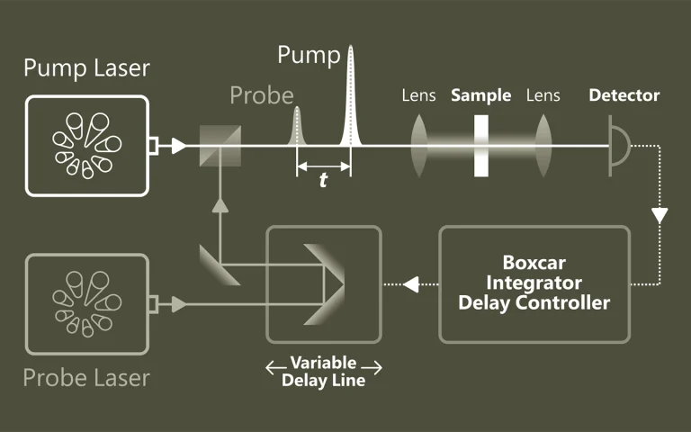
Pumpen. Proben. Verstehen.
Die Pump-Probe-Spektroskopie ermöglicht einen tiefen Einblick in ultraschnelle Prozesse auf atomarer und molekularer Ebene. Mithilfe von gepulsten Lasern lassen sich dynamische Veränderungen mit extrem hoher zeitlicher Auflösung verfolgen. Typische Anwendungen liegen in der Untersuchung von Ladungsträgerdynamik in Halbleitern, der Analyse von Elektronentransfer in chemischen Reaktionen sowie der Erforschung lichtinduzierter Phasenübergänge in Materialien.
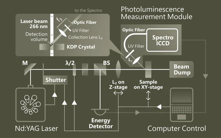
Emission, die Einblicke gibt
Die Photolumineszenz-Spektroskopie (PL) ist eine berührungslose Methode zur Untersuchung elektronischer und struktureller Eigenschaften von Materialien. Dabei wird das Material durch Licht angeregt und die emittierte Strahlung analysiert. Diese spektrale Information gibt Aufschluss über Bandlücken, Defekte, Dotierungen oder Quantenstruktur. PL ist besonders wertvoll für Halbleiter, Nanostrukturen und optoelektronische Materialien
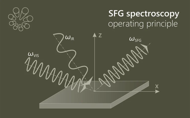
Die Summe macht den Unterschied
Die SFG-Spektroskopie (Sum-Frequency Generation) ist eine nichtlineare optische Methode zur Untersuchung von Molekülen an Oberflächen und Grenzflächen (z. B. Fest-Flüssig, Fest-Gas). Zwei Laserstrahlen – ein sichtbarer und ein infraroter – erzeugen durch Überlagerung ein Summenfrequenzsignal, das nur an nicht-zentrosymmetrischen Strukturen entsteht. Durch Abstimmen des IR-Strahls auf molekulare Schwingungen liefert SFG selektive und …
Anwendungen
Wissenschafliche Veröffentlichungen
Structure Determination of Hen Egg-White Lysozyme Aggregates Adsorbed to Lipid/Water and Air/Water Interfaces
S. Strazdaite, E. Navakauskas, J. Kirschner, T. Sneideris, and G. Niaura, Langmuir 36 (17), 4766-4775 (2020). DOI: 10.1021/acs.langmuir.9b03826.
We use vibrational sum-frequency generation (VSFG) spectroscopy to study the structure of hen egg-white lysozyme (HEWL) aggregates adsorbed to DOPG/D2O and air/D2O interfaces. We find that aggregates with a parallel and antiparallel β-sheet structure together with smaller unordered aggregates and a denaturated protein are adsorbed to both interfaces. We demonstrate that to retrieve this information, fitting of the VSFG spectra is essential. The number of bands contributing to the VSFG spectrum might be misinterpreted, due to interference between peaks with opposite orientation and a nonresonant background. Our study identified hydrophobicity as the main driving force for adsorption to the air/D2O interface. Adsorption to the DOPG/D2O interface is also influenced by hydrophobic interaction; however, electrostatic interaction between the charged protein’s groups and the lipid’s headgroups has the most significant effect on the adsorption. We find that the intensity of the VSFG spectrum at the DOPG/D2O interface is strongly enhanced by varying the pH of the solution. We show that this change is not due to a change of lysozyme’s and its aggregates’ charge but due to dipole reorientation at the DOPG/D2O interface. This finding suggests that extra care must be taken when interpreting the VSFG spectrum of proteins adsorbed at the lipid/water interface.
How nature covers its bases
S. Boldissar, and M. S. de Vries, Phys. Chem. Chem. Phys. 20, 9701-9716 (2018). DOI: 10.1039/C8CP01236A.
The response of DNA and RNA bases to ultraviolet (UV) radiation has been receiving increasing attention for a number of important reasons: (i) the selection of the building blocks of life on an early earth may have been mediated by UV photochemistry, (ii) radiative damage of DNA depends critically on its photochemical properties, and (iii) the processes involved are quite general and play a role in more biomolecules as well as in other compounds. A growing number of groups worldwide have been studying the photochemistry of nucleobases and their derivatives. Here we focus on gas phase studies, which (i) reveal intrinsic properties distinct from effects from the molecular environment, (ii) allow for the most detailed comparison with the highest levels of computational theory, and (iii) provide isomeric selectivity. From the work so far a picture is emerging of rapid decay pathways following UV excitation. The main understanding, which is now well established, is that canonical nucleobases, when absorbing UV radiation, tend to eliminate the resulting electronic excitation by internal conversion (IC) to the electronic ground state in picoseconds or less. The availability of this rapid “safe” de-excitation pathway turns out to depend exquisitely on molecular structure. The canonical DNA and RNA bases are generally short-lived in the excited state, and thus UV protected. Many closely related compounds are longer lived, and thus more prone to other, potentially harmful, photochemical processes. It is this structure dependence that suggests a mechanism for the chemical selection of the building blocks of life on an early earth. However, the picture is far from complete and many new questions now arise.
Vibrational fingerprint of localized excitons in a two-dimensional metal-organic crystal
M. Corva, A. Ferrari, M. Rinaldi, Z. Feng, M. Roiaz, C. Rameshan et al., Nature Communications , 4703 (2018). DOI: 10.1038/s41467-018-07190-1.
Long-lived excitons formed upon visible light absorption play an essential role in photovoltaics, photocatalysis, and even in high-density information storage. Here, we describe a self-assembled two-dimensional metal-organic crystal, composed of graphene-supported macrocycles, each hosting a single FeN4 center, where a single carbon monoxide molecule can adsorb. In this heme-like biomimetic model system, excitons are generated by visible laser light upon a spin transition associated with the layer 2D crystallinity, and are simultaneously detected via the carbon monoxide ligand stretching mode at room temperature and near-ambient pressure. The proposed mechanism is supported by the results of infrared and time-resolved pump-probe spectroscopies, and by ab initio theoretical methods, opening a path towards the handling of exciton dynamics on 2D biomimetic crystals.