PGx01
Ultra-wide tunable picosecond OPA with high pulse energy
These OPAs are an excellent choice for researchers who require a tunable coherent light source with picosecond pulses with high pulse energy from 193nm in the UV to 16 µm in the mid-IR.
Developed and manufactured by:
Features
- Ultra-wide spectral range from 193 to 16,000 nm
- High peak power (> 50 MW), ideal for non-linear spectroscopy
- Narrow line width < 6 cm-¹ (for UV < 9 cm-1)
- Motorized, software-controlled wavelength tuning in the range of 193-2,300 nm or 2,300-16,000 nm
Applications
- Nonlinear spectroscopy: vibrational SFG, surface SH, Z-scan
- Pump-probe experiments
- Laser-induced fluorescence (LIF)
Applications
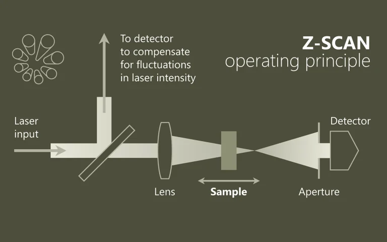
At the focal point of non-linearity
In the field of nonlinear optics, the n2 index (Kerr nonlinearity) or the nonlinear absorption coefficient Δα, for example, are determined using Z-scan. Both the real part and the imaginary part of the nonlinear refractive index are measured using the “open” and “closed” methods.
Pumps. Samples. Understanding.
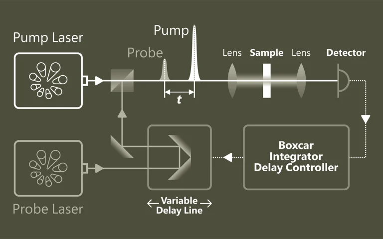
Pump-probe spectroscopy provides a deep insight into ultrafast processes at the atomic and molecular level. With the help of pulsed lasers, dynamic changes can be tracked with extremely high temporal resolution. Typical applications include the investigation of charge carrier dynamics in semiconductors, the analysis of electron transfer in chemical reactions and the study of light-induced phase transitions in materials.
Emission that provides insights
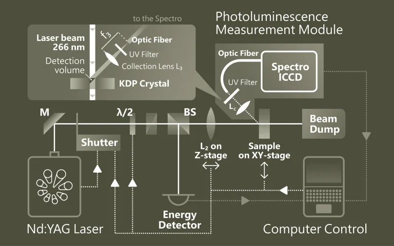
Photoluminescence spectroscopy (PL) is a non-contact method for investigating the electronic and structural properties of materials. The material is excited by light and the emitted radiation is analyzed. This spectral information provides information about band gaps, defects, doping or quantum structure. PL is particularly valuable for semiconductors, nanostructures and optoelectronic materials
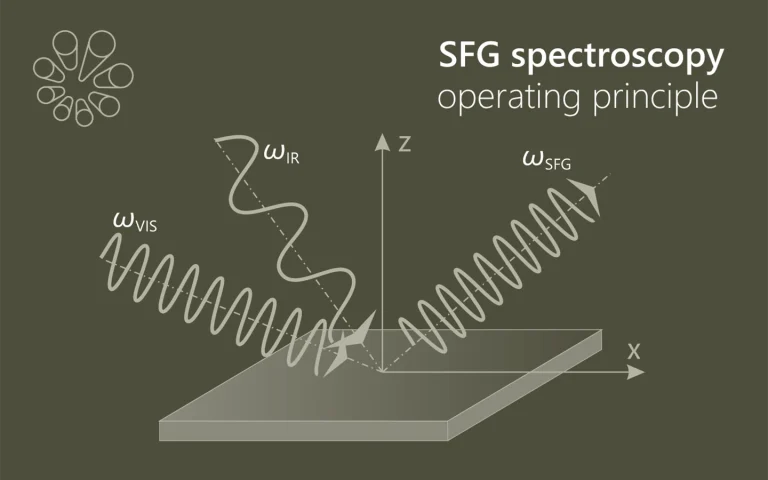
The sum makes the difference
SFG spectroscopy (Sum-Frequency Generation) is a non-linear optical method for investigating molecules on surfaces and interfaces (e.g. solid-liquid, solid-gas). Two laser beams – one visible and one infrared – are superimposed to generate a sum frequency signal that only occurs on non-centrosymmetric structures. By tuning the IR beam to molecular vibrations, SFG provides selective and …
Applications
Scientific publications
Structure Determination of Hen Egg-White Lysozyme Aggregates Adsorbed to Lipid/Water and Air/Water Interfaces
S. Strazdaite, E. Navakauskas, J. Kirschner, T. Sneideris, and G. Niaura, Langmuir 36 (17), 4766-4775 (2020). DOI: 10.1021/acs.langmuir.9b03826.
We use vibrational sum-frequency generation (VSFG) spectroscopy to study the structure of hen egg-white lysozyme (HEWL) aggregates adsorbed to DOPG/D2Oand air/D2Ointerfaces. We find that aggregates with a parallel and antiparallel β-sheet structure together with smaller unordered aggregates and a denaturated protein are adsorbed to both interfaces. We demonstrate that to retrieve this information, fitting of the VSFG spectra is essential. The number of bands contributing to the VSFG spectrum might be misinterpreted, due to interference between peaks with opposite orientation and a nonresonant background. Our study identified hydrophobicity as the main driving force for adsorption to the air/D2Ointerface. Adsorption to the DOPG/D2Ointerface is also influenced by hydrophobic interaction; however, electrostatic interaction between the charged protein’s groups and the lipid’s headgroups has the most significant effect on the adsorption. We find that the intensity of the VSFG spectrum at the DOPG/D2Ointerface is strongly enhanced by varying the pH of the solution. We show that this change is not due to a change of lysozyme’s and its aggregates’ charge but due to dipole reorientation at the DOPG/D2Ointerface. This finding suggests that extra care must be taken when interpreting the VSFG spectrum of proteins adsorbed at the lipid/water interface.
How nature covers its bases
S. Boldissar, and M. S. de Vries, Phys. Chem. Chem. Phys. 20, 9701-9716 (2018). DOI: 10.1039/C8CP01236A.
The response of DNA and RNA bases to ultraviolet (UV) radiation has been receiving increasing attention for a number of important reasons: (i) the selection of the building blocks of life on an early earth may have been mediated by UV photochemistry, (ii) radiative damage of DNA depends critically on its photochemical properties, and (iii) the processes involved are quite general and play a role in more biomolecules as well as in other compounds. A growing number of groups worldwide have been studying the photochemistry of nucleobases and their derivatives. Here we focus on gas phase studies, which (i) reveal intrinsic properties distinct from effects from the molecular environment, (ii) allow for the most detailed comparison with the highest levels of computational theory, and (iii) provide isomeric selectivity. From the work so far a picture is emerging of rapid decay pathways following UV excitation. The main understanding, which is now well established, is that canonical nucleobases, when absorbing UV radiation, tend to eliminate the resulting electronic excitation by internal conversion (IC) to the electronic ground state in picoseconds or less. The availability of this rapid “safe” de-excitation pathway turns out to depend exquisitely on molecular structure. The canonical DNA and RNA bases are generally short-lived in the excited state, and thus UV protected. Many closely related compounds are longer lived, and thus more prone to other, potentially harmful, photochemical processes. It is this structure dependence that suggests a mechanism for the chemical selection of the building blocks of life on an early earth. However, the picture is far from complete and many new questions now arise.
Vibrational fingerprint of localized excitons in a two-dimensional metal-organic crystal
M. Corva, A. Ferrari, M. Rinaldi, Z. Feng, M. Roiaz, C. Rameshan et al, Nature Communications , 4703 (2018). DOI: 10.1038/s41467-018-07190-1.
Long-lived excitons formed upon visible light absorption play an essential role in photovoltaics, photocatalysis, and even in high-density information storage. Here, we describe a self-assembled two-dimensional metal-organic crystal, composed of graphene-supported macrocycles, each hosting a single FeN4 center, where a single carbon monoxide molecule can adsorb. In this heme-like biomimetic model system, excitons are generated by visible laser light upon a spin transition associated with the layer 2D crystallinity, and are simultaneously detected via the carbon monoxide ligand stretching mode at room temperature and near-ambient pressure. The proposed mechanism is supported by the results of infrared and time-resolved pump-probe spectroscopies, and by ab initio theoretical methods, opening a path towards the handling of exciton dynamics on 2D biomimetic crystals.