NT240
Abstimmbarer Diodengepumpter Laser mit kHz Repetitionsrate
Der NT240 bietet motorisiertes, lückenloses Wellenlängen-Tuning von 210 bis 2600 nm – aus nur einem Gehäuse. Mit 1000 Hz Wiederholrate ist er ein vielseitiges Werkzeug für Anwendungen wie LIF, Pump-Probe, Photolyse, Photobiologie, Metrologie und Fernerkundung.
Entwickelt und hergestellt von:
Features
- Zwei Jahre Gewährleistung
- Integriert DPSS-Pumplaser und OPO in einem Gehäuse
- Motorisiertes, lückenloses Wellenlängen-Tuning von 210 bis 2600 nm*
- 1000 Hz Pulswiederholrate
- Über 60 µJ Ausgangspulsenergie im UV
- Linienbreite < 5 cm⁻¹
- 3–6 ns Pulsdauer
- Fernsteuerung über Tastenfeld oder PC
- Optional separater Ausgang für OPO-Pumpstrahl: 355 nm, 532 nm oder 1064 nm
Automatischer Wellenlängenscan ist programmierbar
Anwendungen
- Laserinduzierte
- Fluoreszenzspektroskopie
- Pump-Probe-Spektroskopie
- Nichtlineare Spektroskopie
- Zeitaufgelöste
- Spektroskopie
- Photobiologie
- Fernerkundung
Anwendungen
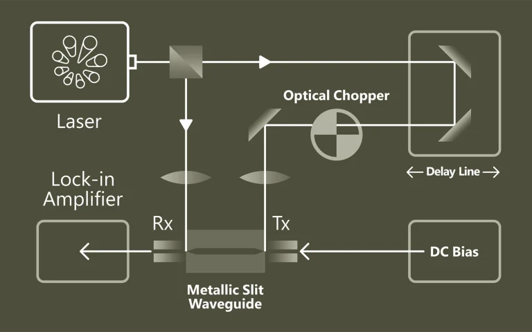
Ladung unter Kontrolle
Zeitaufgelöste Photoleitfähigkeit (TRMC) ermöglicht kontaktfreie Analyse von Ladungsträgerdichte, Transport- und Rekombinationsparametern in Materialien wie organischen Halbleitern, Farbstoffen oder Kompositen. Sie liefert wichtige Erkenntnisse über lichtinduzierte Prozesse abhängig von Photonenenergie, Intensität, Temperatur und Zeitverlauf. Heute dient TRMC als präzises Diagnosewerkzeug zur Charakterisierung neuer elektronischer Materialien und Bauelemente – etwa für Tinten, LEDs, Katalyse oder Photovoltaik.
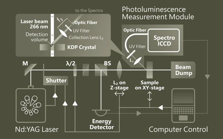
Emission, die Einblicke gibt
Die Photolumineszenz-Spektroskopie (PL) ist eine berührungslose Methode zur Untersuchung elektronischer und struktureller Eigenschaften von Materialien. Dabei wird das Material durch Licht angeregt und die emittierte Strahlung analysiert. Diese spektrale Information gibt Aufschluss über Bandlücken, Defekte, Dotierungen oder Quantenstruktur. PL ist besonders wertvoll für Halbleiter, Nanostrukturen und optoelektronische Materialien
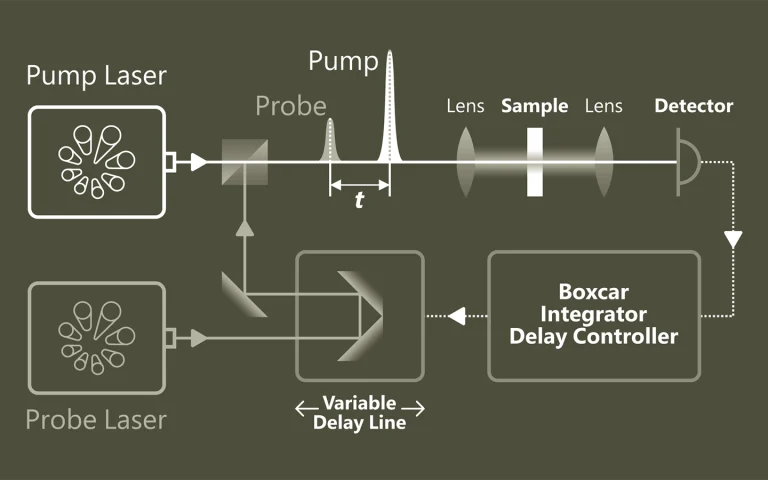
Pumpen. Proben. Verstehen
Die Pump-Probe-Spektroskopie ermöglicht einen tiefen Einblick in ultraschnelle Prozesse auf atomarer und molekularer Ebene. Mithilfe von gepulsten Lasern lassen sich dynamische Veränderungen mit extrem hoher zeitlicher Auflösung verfolgen. Typische Anwendungen liegen in der Untersuchung von Ladungsträgerdynamik in Halbleitern, der Analyse von Elektronentransfer in chemischen Reaktionen sowie der Erforschung lichtinduzierter Phasenübergänge in Materialien.
Wenn Licht Moleküle trennt
Photolyse beschreibt die lichtinduzierte Spaltung chemischer Verbindungen und spielt eine zentrale Rolle in der Photosynthese durch Wasserspaltung. Die Laser-Flash-Photolyse nutzt kurze, intensive Laserpulse (ns–fs), um kurzlebige Zwischenprodukte wie Radikale oder angeregte Zustände zu erzeugen und deren zeitlich aufgelöste Absorption zu beobachten. Probe-Pulse erfassen Reaktionsverläufe. Der Einsatz von OPOs erweitert die Möglichkeiten spektroskopischer Untersuchungen deutlich.
Anwendungen
Wissenschafliche Veröffentlichungen
Contrast agent enhanced multimodal photoacoustic microscopy and optical coherence tomography for imaging of rabbit choroidal and retinal vessels in vivo
V. P. Nguyen, Y. Li, W. Qian, B. Liu, C. Tian, W. Zhang et al., Scientific Reports 9 (1), 5945 (2019). DOI: 10.1038/s41598-019-42324-5.
Multimodal imaging with photoacoustic microscopy (PAM) and optical coherence tomography (OCT) can be an effective method to evaluate the choroidal and retinal microvasculature. To improve the efficiency for visualizing capillaries, colloidal gold nanoparticles (AuNPs) have been applied as a multimodal contrast agent for both OCT and PAM imaging by taking advantage of the strong optical scattering and the strong optical absorption of AuNPs due to their surface plasmon resonance. Ultra-pure AuNPs were fabricated by femtosecond laser ablation, capped with polyethylene glycol (PEG), and administered to 13 New Zealand white rabbits and 3 Dutch Belted pigmented rabbits. The synthesized PEG-AuNPs (20.0 ± 1.5 nm) were demonstrated to be excellent contrast agents for PAM and OCT, and do not demonstrate cytotoxicity to bovine retinal endothelial cells in cell studies. The image signal from the retinal and choroidal vessels in living rabbits was enhanced by up to 82% for PAM and up to 45% for OCT, respectively, by the administered PEG-AuNPs, which enables detection of individual blood vessels by both imaging modalities. The biodistribution study demonstrated the AuNP accumulated primarily in the liver and spleen. Histology and TUNEL staining did not indicate cell injury or death in the lung, liver, kidney, spleen, heart, or eyes up to seven days after AuNP administration. PEG-AuNPs offer an efficient and safe contrast agent for multimodal ocular imaging to achieve improved characterization of microvasculature.
High-resolution multimodal photoacoustic microscopy and optical coherence tomography image-guided laser induced branch retinal vein occlusion in living rabbits
V. P. Nguyen, Y. Li, W. Zhang, X. Wang, and Y. M. Paulus, Scientific reports 9 (1), 10560 (2019). DOI: 10.1038/s41598-019-47062-2.
Joint high-resolution multimodal photoacoustic microscopy (PAM) and optical coherence tomography (OCT) was developed to improve the efficiency for visualizing newly developed retinal neovascularization (RNV) and to monitor the dynamic changes of retinal vein occlusion (RVO) in living rabbits. The RNV and RVO models were created in New Zealand rabbits by Rose Bengal laser-induced RVO. Dual modalities imaging equipment, including color fundus photography, fluorescein angiography (FA), OCT, and PAM, was used to image and assess the changes of retinal vasculature. In vivo experimental results exhibited that not only the treatment boundaries and the position of the occluded vasculature but also the structure of individual RNV were markedly observed using PAM platform with great resolution and high image contrast. The laser light energy of 80 nJ was used to induce photoacoustic signal, which is approximately half the energy of the American National Standards Institute safety limit. A cross-sectional structure of RNV was identified with the OCT modality. Furthermore, vibrant transformations in the RNV and the retinal morphology were examined at different times after laser occlusion: days 4, 28, 35, 49, and 90. PAM revealed high contrast and high resolution vascular imaging of the retina and choroid with amplified penetration depth. Through the present custom-built imaging system, both RNV and RVO can be reconstructed and observed in two and three dimensions. A unique dual modality A unique dual modality PAM and OCT can help precisely visualize and distinguish individual microvessels, microvessel depth, and the surrounding anatomy. Thus, the proposed multimodal ocular imaging platform may offer a potential equipment to enhance classification of microvasculature in a reliable and proficient manner in larger rabbit eyes.
High-resolution, high-contrast mid-infrared imaging of fresh biological samples with ultraviolet-localized photoacoustic microscopy
J. Shi, T. T. W. Wong, Y. He, L. Li, R. Zhang, C. S. Yung et al., Nature Photonics 13 (9), 609-615 (2019). DOI: 10.1038/s41566-019-0441-3.
Mid-infrared (MIR) microscopy provides rich chemical and structural information about biological samples, without staining. Conventionally, the long MIR wavelength severely limits the lateral resolution owing to optical diffraction; moreover, the strong MIR absorption of water ubiquitous in fresh biological samples results in high background and low contrast. To overcome these limitations, we propose a method that employs photoacoustic detection highly localized with a pulsed ultraviolet laser on the basis of the Grüneisen relaxation effect. For cultured cells, our method achieves water-background suppressed MIR imaging of lipids and proteins at ultraviolet resolution, at least an order of magnitude finer than the MIR diffraction limits. Label-free histology using this method is also demonstrated in thick brain slices. Our approach provides convenient high-resolution and high-contrast MIR imaging, which can benefit the diagnosis of fresh biological samples.