NT240
Tunable diode-pumped laser with kHz repetition rate
The NT240 offers motorized, gapless wavelength tuning from 210 to 2600 nm – from just one housing. With 1000 Hz repetition rate, it is a versatile tool for applications such as LIF, pump-probe, photolysis, photobiology, metrology and remote sensing.
Developed and manufactured by:
Features
- Two-year warranty
- Integrates DPSS pump laser and OPO in one housing
- Motorized, seamless wavelength tuning from 210 to 2600 nm*
- 1000 Hz pulse repetition rate
- Over 60 µJ output pulse energy in the UV
- Line width <5 cm-¹
- 3-6 ns Pulse duration
- Remote control via keypad or PC
- Optional separate output for OPO pump beam: 355 nm, 532 nm or 1064 nm
Automatic wavelength scan is programmable
Applications
- Laser-induced
- Fluorescence spectroscopy
- Pump-probe spectroscopy
- Nonlinear spectroscopy
- Time-resolved
- Spectroscopy
- Photobiology
- Remote sensing
Applications
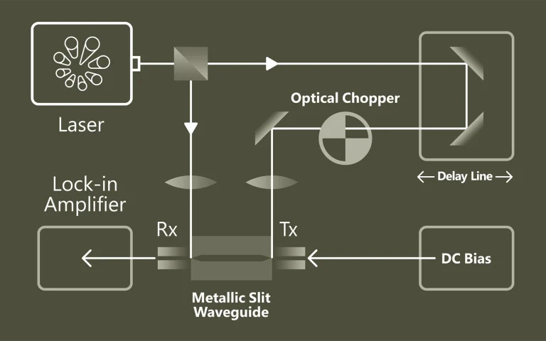
Load under control
Time-resolved photoconductivity (TRMC) enables contact-free analysis of charge carrier density, transport and recombination parameters in materials such as organic semiconductors, dyes or composites. It provides important insights into light-induced processes depending on photon energy, intensity, temperature and time course. Today, TRMC serves as a precise diagnostic tool for characterizing new electronic materials and components – for example for inks, LEDs, catalysis or photovoltaics.
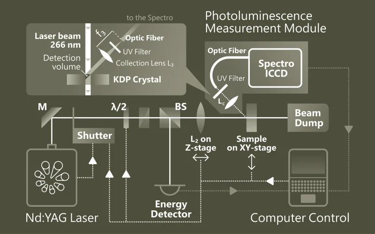
Emission that provides insights
Photoluminescence spectroscopy (PL) is a non-contact method for investigating the electronic and structural properties of materials. The material is excited by light and the emitted radiation is analyzed. This spectral information provides information about band gaps, defects, doping or quantum structure. PL is particularly valuable for semiconductors, nanostructures and optoelectronic materials
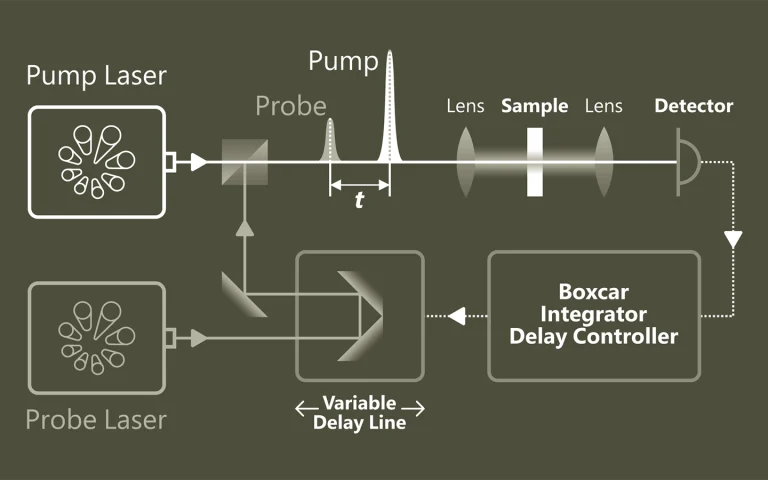
Pumps. Samples. Understanding
Pump-probe spectroscopy provides a deep insight into ultrafast processes at the atomic and molecular level. With the help of pulsed lasers, dynamic changes can be tracked with extremely high temporal resolution. Typical applications are the investigation of charge carrier dynamics in semiconductors, the analysis of electron transfer in chemical reactions and the study of light-induced phase transitions in materials.
When light separates molecules
Photolysis describes the light-induced splitting of chemical compounds and plays a central role in photosynthesis through water splitting. Laser flash photolysis uses short, intense laser pulses (ns-fs) to generate short-lived intermediates such as radicals or excited states and to observe their temporally resolved absorption. Probe pulses record reaction processes. The use of OPOs significantly expands the possibilities of spectroscopic investigations.
Applications
Scientific publications
Contrast agent enhanced multimodal photoacoustic microscopy and optical coherence tomography for imaging of rabbit choroidal and retinal vessels in vivo
V. P. Nguyen, Y. Li, W. Qian, B. Liu, C. Tian, W. Zhang et al, Scientific Reports 9 (1), 5945 (2019). DOI: 10.1038/s41598-019-42324-5.
Multimodal imaging with photoacoustic microscopy (PAM) and optical coherence tomography (OCT) can be an effective method to evaluate the choroidal and retinal microvasculature. To improve the efficiency for visualizing capillaries, colloidal gold nanoparticles (AuNPs) have been applied as a multimodal contrast agent for both OCT and PAM imaging by taking advantage of the strong optical scattering and the strong optical absorption of AuNPs due to their surface plasmon resonance. Ultra-pure AuNPs were fabricated by femtosecond laser ablation, capped with polyethylene glycol (PEG), and administered to 13 New Zealand white rabbits and 3 Dutch Belted pigmented rabbits. The synthesized PEG-AuNPs (20.0 ± 1.5 nm) were demonstrated to be excellent contrast agents for PAM and OCT, and do not demonstrate cytotoxicity to bovine retinal endothelial cells in cell studies. The image signal from the retinal and choroidal vessels in living rabbits was enhanced by up to 82% for PAM and up to 45% for OCT, respectively, by the administered PEG-AuNPs, which enables detection of individual blood vessels by both imaging modalities. The biodistribution study demonstrated the AuNP accumulated primarily in the liver and spleen. Histology and TUNEL staining did not indicate cell injury or death in the lung, liver, kidney, spleen, heart, or eyes up to seven days after AuNP administration. PEG-AuNPs offer an efficient and safe contrast agent for multimodal ocular imaging to achieve improved characterization of microvasculature.
High-resolution multimodal photoacoustic microscopy and optical coherence tomography image-guided laser induced branch retinal vein occlusion in living rabbits
V. P. Nguyen, Y. Li, W. Zhang, X. Wang, and Y. M. Paulus, Scientific reports 9 (1), 10560 (2019). DOI: 10.1038/s41598-019-47062-2.
Joint high-resolution multimodal photoacoustic microscopy (PAM) and optical coherence tomography (OCT) was developed to improve the efficiency for visualizing newly developed retinal neovascularization (RNV) and to monitor the dynamic changes of retinal vein occlusion (RVO) in living rabbits. The RNV and RVO models were created in New Zealand rabbits by Rose Bengal laser-induced RVO. Dual modalities imaging equipment, including color fundus photography, fluorescein angiography (FA), OCT, and PAM, was used to image and assess the changes of retinal vasculature. In vivo experimental results exhibited that not only the treatment boundaries and the position of the occluded vasculature but also the structure of individual RNV were markedly observed using PAM platform with great resolution and high image contrast. The laser light energy of 80 nJ was used to induce photoacoustic signal, which is approximately half the energy of the American National Standards Institute safety limit. A cross-sectional structure of RNV was identified with the OCT modality. Furthermore, vibrant transformations in the RNV and the retinal morphology were examined at different times after laser occlusion: days 4, 28, 35, 49, and 90. PAM revealed high contrast and high resolution vascular imaging of the retina and choroid with amplified penetration depth. Through the present custom-built imaging system, both RNV and RVO can be reconstructed and observed in two and three dimensions. A unique dual modality A unique dual modality PAM and OCT can help precisely visualize and distinguish individual microvessels, microvessel depth, and the surrounding anatomy. Thus, the proposed multimodal ocular imaging platform may offer a potential equipment to enhance classification of microvasculature in a reliable and proficient manner in larger rabbit eyes.
High-resolution, high-contrast mid-infrared imaging of fresh biological samples with ultraviolet-localized photoacoustic microscopy
J. Shi, T. T. W. Wong, Y. He, L. Li, R. Zhang, C. S. Yung et al, Nature Photonics 13 (9), 609-615 (2019). DOI: 10.1038/s41566-019-0441-3.
Mid-infrared (MIR) microscopy provides rich chemical and structural information about biological samples, without staining. Conventionally, the long MIR wavelength severely limits the lateral resolution owing to optical diffraction; moreover, the strong MIR absorption of water ubiquitous in fresh biological samples results in high background and low contrast. To overcome these limitations, we propose a method that employs photoacoustic detection highly localized with a pulsed ultraviolet laser on the basis of the Grüneisen relaxation effect. For cultured cells, our method achieves water-background suppressed MIR imaging of lipids and proteins at ultraviolet resolution, at least an order of magnitude finer than the MIR diffraction limits. Label-free histology using this method is also demonstrated in thick brain slices. Our approach provides convenient high-resolution and high-contrast MIR imaging, which can benefit the diagnosis of fresh biological samples.