PhotoSonus X
DPSS high-power laser with adjustable wavelength
The combination of high output energy, wide wavelength tuning range, repetition rates of up to 100 Hz and fast wavelength change makes this laser source particularly suitable for photoacoustic imaging. It enables the acquisition of high-resolution image data at a high acquisition rate.
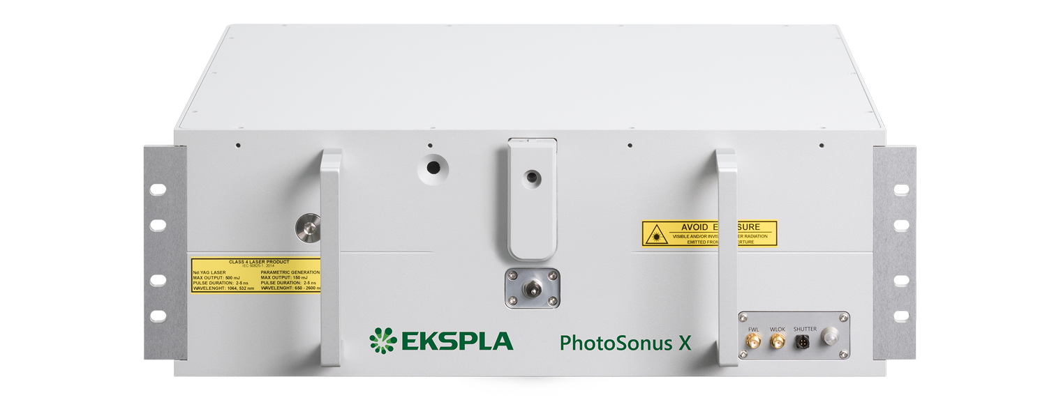
Developed and manufactured by:
Features
- Ultra-wide signal tuning range from 650 to 1300 nm
- Fully motorized wavelength adjustment
- Fast wavelength change
- Externally triggerable
- High pulse energy of up to 90 mJ from the OPO
- Pulse repetition rate of 100 Hz or 50 Hz
- Ready for certification
- Low-noise operation with <60 dB
- Integrated DPSS pump laser and OPO in one housing
- Light extraction via fiber bundles or individual fibers
- Signal and idler beam via the same output (optional)
- Integrated energy meter (optional)
- Electromechanical output shutter with integrated laser self-test function
Highlights
- Pulse repetition rate of 100Hz or 50Hz
- Integrated DPSS pump laser and OPO in one housing
- Ready for certification
- Low-noise operation with <60 dB
Applications
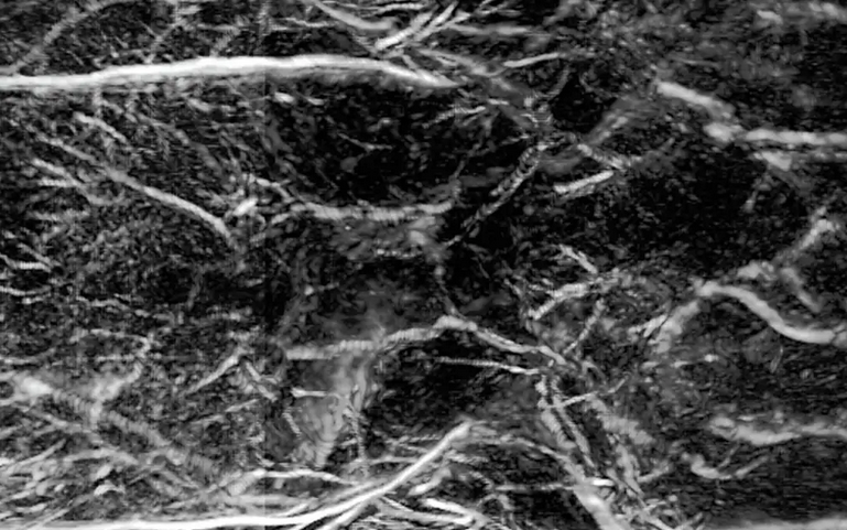
Hearing with light - seeing with sound
Laser-based photoacoustic imaging is an innovative method that combines light and sound to generate high-resolution images of biological tissue. Tissue is irradiated with short, usually spectrally tunable laser pulses. These lead to minimal local heating and thus generate ultrasonic waves, which are measured and converted into images.
When light separates molecules
Photolysis describes the light-induced splitting of chemical compounds and plays a central role in photosynthesis through water splitting. Laser flash photolysis uses short, intense laser pulses (ns-fs) to generate short-lived intermediates such as radicals or excited states and to observe their temporally resolved absorption. Probe pulses record reaction processes. The use of OPOs significantly expands the possibilities of spectroscopic investigations.
Applications
Scientific publications
A fast all-optical 3D photoacoustic scanner for clinical vascular imaging
N. T. Huynh, E. Zhang, O. Francies, F. Kuklis, T. Allen, J. Zhu et al, Nature Biomedical Engineering (2024). DOI: 10.1038/s41551-024-01247-x.
The clinical assessment of microvascular pathologies (in diabetes and in inflammatory skin diseases, for example) requires the visualization of superficial vascular anatomy. Photoacoustic tomography (PAT) scanners based on an all-optical Fabry-Perot ultrasound sensor can provide highly detailed 3D microvascular images, but minutes-long acquisition times have precluded their clinical use. Here we show that scan times can be reduced to a few seconds and even hundreds of milliseconds by parallelizing the optical architecture of the sensor readout, by using excitation lasers with high pulse-repetition frequencies and by exploiting compressed sensing. A PAT scanner with such fast acquisition minimizes motion-related artefacts and allows for the volumetric visualization of individual arterioles, venules, venous valves and millimetre-scale arteries and veins to depths approaching 15 mm, as well as for dynamic 3D images of time-varying tissue perfusion and other haemodynamic events. In exploratory case studies, we used the scanner to visualize and quantify microvascular changes associated with peripheral vascular disease, skin inflammation and rheumatoid arthritis. Fast all-optical PAT may prove useful in cardiovascular medicine, oncology, dermatology and rheumatology.
Theoretical and experimental comparison of the performance of gold, titanium, and platinum nanodiscs as contrast agents for photoacoustic imaging
J. Wi, J. Kim, M. Y. Kim, S. Choi, H. J. Jung, C. Kim et al, RSC Adv. 13, 9441-9447 (2023). DOI: 10.1039/D3RA00795B.
Exogenous contrast agents in photoacoustic imaging help improve spatial resolution and penetration depth and enable targeted molecular imaging. To screen efficient photoacoustic signaling materials as contrast agents, we propose a light absorption-weighted figure of merit (FOM) that can be calculated using material data from the literature and numerically simulated light absorption cross-sections. The calculated light absorption-weighted FOM shows that a Ti nanodisc has a photoacoustic conversion performance similar to that of an Au nanodisc and better than that of a Pt nanodisc. The photoacoustic imaging results of Ti, Au, and Pt nanodiscs, which are physically synthesized with identical shapes and dimensions, experimentally demonstrated that the Ti nanodisc could be a highly efficient contrast agent.
Utilizing nanosecond sources in diffuse optical tomography
M. Mozumder, J. Leskinen, and T. Tarvainen, Measurement Science and Technology 34 (2), 025901 (2022). DOI: 10.1088/1361-6501/ac9e11.
Diffuse optical tomography (DOT) use near-infrared light for imaging optical properties of biological tissues. Time-domain (TD) DOT systems use pulsed lasers and measure time-varying temporal point spread function (TPSF), carrying information from both superficial and deep layers of imaged target. In this work, feasibility of nanosecond scale light pulses as sources for TD-DOT is studied. Nanosecond sources enable using relatively robust measurement setups with standard analogue-to-digital converter waveform digitizers, such as digital oscilloscopes. However, these type of systems have some properties, such as variations in source pulses and limited temporal sampling, that could limit their usage. In this work, these different aspects and possible limitations were studied with simulations and experiments. Simulations showed that information carried by TD data of diffuse medium is on low frequencies. This enables usage of relatively slow response time measurement electronics, and image processing using Fourier-transformed TD data. Furthermore, the temporal sampling in measurements needs to be high enough to capture the TPSF, but this rate can be achieved with standard digital oscilloscopes. It was shown that, although variations in light pulses of nanosecond lasers are larger than those of picosecond sources, these variations do not affect significantly on image quality. Overall, the simulations demonstrated the capability of nanosecond sources to be utilized in TD-DOT in diffuse medium. In this work, a prototype TD-DOT experimental system utilizing a high-energy nanosecond laser was constructed. The system is relatively robust consisting of a nanosecond Nd:YAG laser combined with optical parametric oscillator for light input and optical fibers for guiding the light, and avalanche photodetector and high-bandwidth oscilloscope for TPSF measurements. The system was used in both absolute and difference imaging of two phantoms. The experiments verified that both absorbing and scattering objects can be reconstructed with good accuracy with TD-DOT using a nanosecond laser.