PL2250 Serie
Blitzlampengepumpte Pikosekunden-Nd:YAG-Laser
Die Laser der PL2250-Serie sind modengekoppelte Pikosekunden-Nd:YAG-Laser, die hochenergetische Pulse mit bis zu 100 mJ bei einer Pulswiederholrate von 20 Hz erzeugen. Sie zeichnen sich durch ein kosteneffizientes Design aus, das gleichzeitig eine hohe Zuverlässigkeit gewährleistet und die Betriebs- sowie Wartungskosten reduziert.

Entwickelt und hergestellt von:
Features
- Hermetisch versiegelter DPSS-Masteroszillator
- Diodengepumpter regenerativer Verstärker
- Blitzlampengepumpter Leistungsverstärker mit bis zu 100 mJ Pulsenergie bei 1064 nm
- Pulsdauer von 30 ps (optional 20 ps)
- Hervorragende Pulsdauerkonstanz
- Wiederholrate bis zu 20 Hz
- Triggerpuls für Streak-Kamera mit <10 ps Jitter
- Exzellente Strahllage-Stabilität
- Optional thermostabilisierte Frequenzkonverter für Harmonischen
- Sodtwaregesteuert
- Bedienteil
Anwendungen
- Zeitaufgelöste Fluoreszenz-Spektroskopie (inkl. Streak-Kamera Messungen)
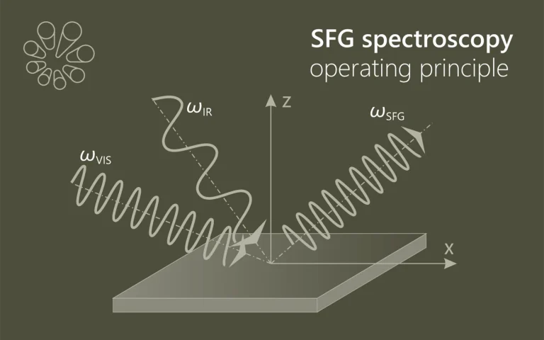
- SFG/SHG-Spektroskopie
- Nichtlineare Spektroskopie
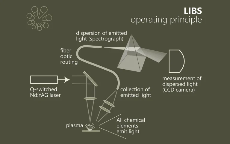
- Laser-Induced Breakdown Spectroscopy (LIBS)
- OPG Pumpen
- Remote laser sensing
- Satellite Ranging
- Weitere Anwendungen im Bereich der Spektroskopie und Nichtlinearen Optik.
Anwendungen
Zerstörung für präzise Erkenntnisse
Die laserinduzierte Plasmaspektroskopie (LIBS) ist eine schnelle, zerstörungsarme Methode zur Bestimmung der Elementzusammensetzung von Materialien. Ein intensiver Laserpuls erzeugt auf der Probenoberfläche ein Plasma, dessen charakteristische Emission analysiert wird. LIBS eignet sich für nahezu alle Materialarten…
Emission, die Einblicke gibt
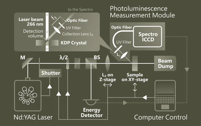
Die Photolumineszenz-Spektroskopie (PL) ist eine berührungslose Methode zur Untersuchung elektronischer und struktureller Eigenschaften von Materialien. Dabei wird das Material durch Licht angeregt und die emittierte Strahlung analysiert. Diese spektrale Information gibt Aufschluss über Bandlücken, Defekte, Dotierungen oder Quantenstruktur. PL ist besonders wertvoll für Halbleiter, Nanostrukturen und optoelektronische Materialien…
Die Summe macht den Unterschied
Die SFG-Spektroskopie (Sum-Frequency Generation) ist eine nichtlineare optische Methode zur Untersuchung von Molekülen an Oberflächen und Grenzflächen (z. B. Fest-Flüssig, Fest-Gas). Zwei Laserstrahlen – ein sichtbarer und ein infraroter – erzeugen durch Überlagerung ein Summenfrequenzsignal, das nur …
Anwendungen
Wissenschafliche Veröffentlichungen
Aggregation states of poly (4-methylpentene-1) at a solid interface
K. Yamamoto, D. Kawaguchi, K. Sasahara, M. Inutsuka, S. Yamamoto, K. Uchida et al., Polymer Journal 51 (2), 247-255 (2019). DOI: 10.1038/s41428-018-0134-7.
A thin film of poly(4-methylpentene-1) (P4MP1) was prepared on a quartz substrate, which was a model system of an interface in filler-reinforced semicrystalline polymer composites. Grazing-incidence wide-angle X-ray diffraction measurements revealed that P4MP1 in the thin film after isothermal crystallization formed a Form I crystal polymorph composed of a tetragonal unit cell with a 72 helix, in which the chain axis was oriented along the direction parallel to the quartz interface. Combining sum-frequency generation vibrational spectroscopy with molecular dynamics simulation enabled us to gain access to the local conformation of P4MP1 chains at the quartz interface and the changes that occurred with isothermal crystallization. Finally, the way in which the initial chain orientation at the substrate interface impacted the crystalline structure in the thin film was discussed.
Soft x-ray emission from laser-produced strontium ions
T. Miyazaki, G. O’Sullivan, and P. Dunne, Journal of Physics B: Atomic, Molecular and Optical Physics 53 (2), 025001 (2019). DOI: 10.1088/1361-6455/ab53be.
Soft x-ray spectra, in the range from 2 nm to 9 nm, were recorded from strontium plasmas formed by pulses from 20 ps, 170 ps and 5.5 ns Nd:YAG lasers operating at the fundamental wavelength of 1064 nm. Features due to 3d–4p and 3d–4f transitions were identified by comparison with spectra from adjacent ions and atomic structure calculations with both the Cowan code and the Flexible Atomic Code. As in the spectra of ions of other elements in the fifth row of the periodic table, resonant lines 3dn–3dn−14p1, 3dn–3dn−14f1 and satellite lines 3dn−14s1–3dn−24s14p1, 3dn−14s1–3dn−24s14f1 of Δn = 1 were observed over the 3.0–8.5 nm region, emitted by 10+ to 19+ ions. These Δn = 1 transitions provide a range of narrow band emission features which may match to specific multi layer combinations for reflective optics in the extreme ultraviolet region of the spectrum.
Intraoperative diagnostics and elimination of residual microtumours with plasmonic nanobubbles
E. Y. Lukianova‑Hleb, Y. Kim, I. Belatsarkouski, A. M. Gillenwater, B. E. O’Neill, and D. O. Lapotko, Nature Nanotechnology 11 (6), 525-532 (2016). DOI: 10.1038/NNANO.2015.343.
Failure of cancer surgery to intraoperatively detect and eliminate microscopic residual disease (MRD) causes lethal recurrence and metastases, and the removal of important normal tissues causes excessive morbidity. Here, we show that a plasmonic nanobubble (PNB), a non-stationary laser pulse-activated nanoevent, intraoperatively detects and eliminates MRD in the surgical bed. PNBs were generated in vivo in head and neck cancer cells by systemically targeting tumours with gold colloids and locally applying near-infrared, low-energy short laser pulses, and were simultaneously detected with an acoustic probe. In mouse models, between 3 and 30 residual cancer cells and MRD (undetectable with current methods) were non-invasively detected up to 4 mm deep in the surgical bed within 1 ms. In resectable MRD, PNB-guided surgery prevented local recurrence and delivered 100% tumour-free survival. In unresectable MRD, PNB nanosurgery improved survival twofold compared with standard surgery. Our results show that PNB-guided surgery and nanosurgery can rapidly and precisely detect and remove MRD in simple intraoperative procedures.