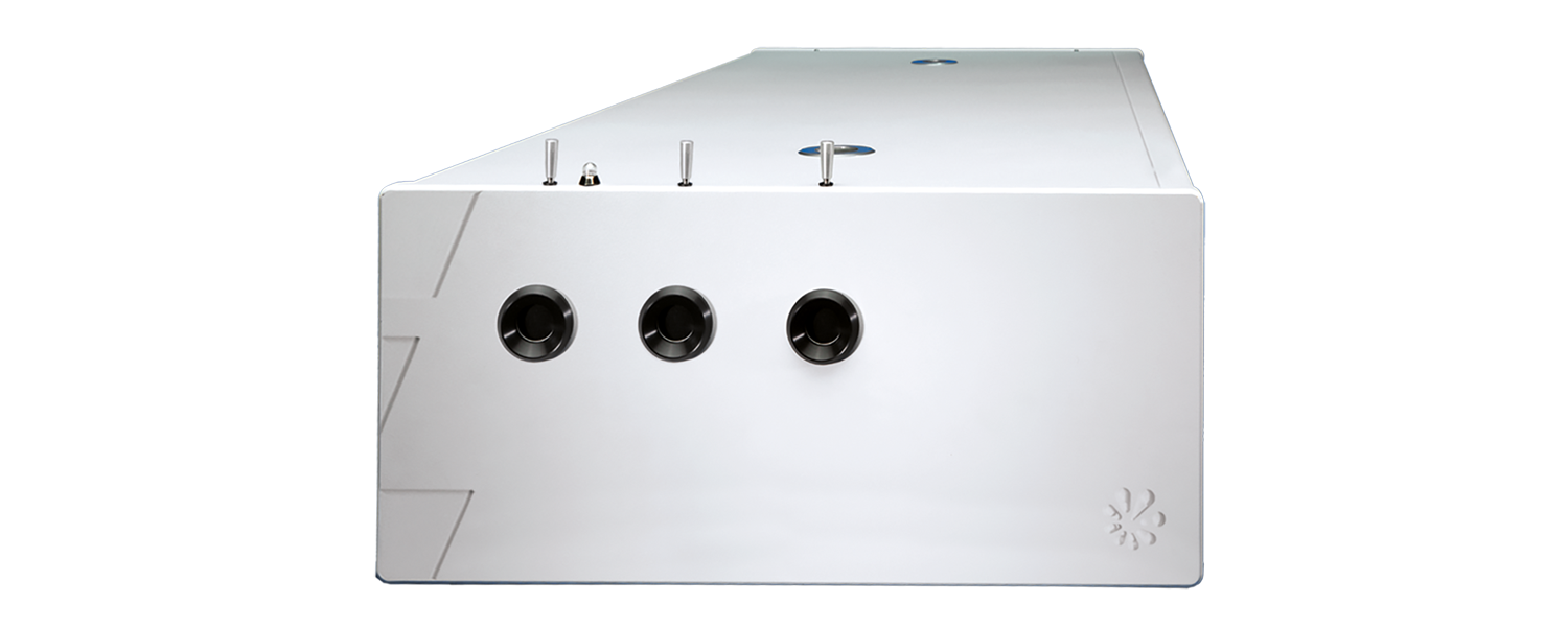
PhotoSonus T
Hochenergetischer abstimmbarer Laser für die photoakustische Bildgebung
Der PhotoSonus T ist die Table-Top Version des hochenergetischen, abstimmbaren Lasers PhotoSonus M. Er bietet hohe Ausgangsenergien zur Abbildung größerer Gewebevolumina im Bereich der photoakustischen Bildgebung.
Entwickelt und hergestellt von:
Features
- Softwaregesteuerte Wellenlängeneinstellung von 330 bis 2600 nm
- Ultrabreiter OPO-Signal-Abstimmbereich von 660 bis 1320 nm
- Bis zu 230 mJ im Bereich 660 – 2600 nm, 35 mJ im Bereich 330 – 660 nm
- Schmale Linienbreite über den gesamten abstimmbaren Bereich
- Pulsdauer von 3–5 ns
- Steuerung über Handgerät oder PC
- Separater Auskopplungsport für 532 nm; optionaler Ausgang für 1064 nm
- Überwachung der OPO-Pumplaserenergie
- Schneller Wellenlängenwechsel innerhalb des gesamten Signal- oder Idlerbereiches
Anwendungen
- Photoakustische Bildgebung
- Blitzphotolyse
- Photobiologie
- Remote Sensing
- Nichtlineare Spektroskopie
Anwendungen
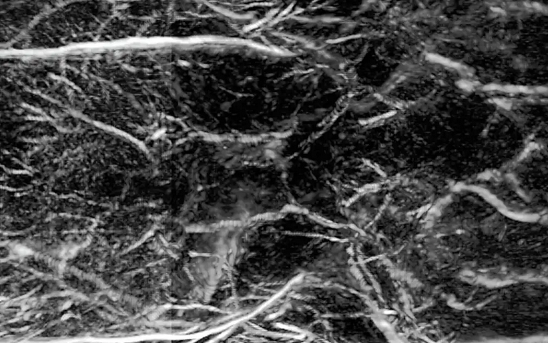
Mit Licht hören – mit Schall sehen
Die laserbasierte photoakustische Bildgebung ist ein innovatives Verfahren, das Licht und Schall kombiniert, um hochauflösende Bilder biologischer Gewebe zu erzeugen. Dabei wird Gewebe mit kurzen, meist spektral abstimmbaren Laserpulsen bestrahlt. Diese führen zu einer minimalen lokalen Erwärmung und erzeugen dadurch Ultraschallwellen, die gemessen und in Bilder umgewandelt werden.
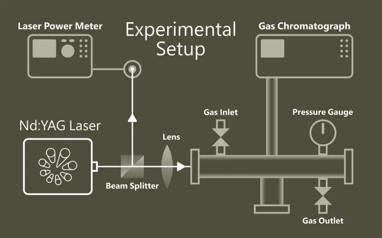
Wenn Licht Moleküle trennt
Photolyse beschreibt die lichtinduzierte Spaltung chemischer Verbindungen und spielt eine zentrale Rolle in der Photosynthese durch Wasserspaltung. Die Laser-Flash-Photolyse nutzt kurze, intensive Laserpulse (ns–fs), um kurzlebige Zwischenprodukte wie Radikale oder angeregte Zustände zu erzeugen und deren zeitlich aufgelöste Absorption zu beobachten. Probe-Pulse erfassen Reaktionsverläufe. Der Einsatz von OPOs erweitert die Möglichkeiten spektroskopischer Untersuchungen deutlich.
Anwendungen
Wissenschafliche Veröffentlichungen
An Investigation of Signal Preprocessing for Photoacoustic Tomography
I. Huen, R. Zhang, R. Bi, X. Li, M. Moothanchery, and M. Olivo, Sensors 23 (1), 510 (2023). DOI: 10.3390/s23010510.
Photoacoustic tomography (PAT) is increasingly being used for high-resolution biological imaging at depth. Signal-to-noise ratios and resolution are the main factors that determine image quality. Various reconstruction algorithms have been proposed and applied to reduce noise and enhance resolution, but the efficacy of signal preprocessing methods which also affect image quality, are seldom discussed. We, therefore, compared common preprocessing techniques, namely bandpass filters, wavelet denoising, empirical mode decomposition, and singular value decomposition. Each was compared with and without accounting for sensor directivity. The denoising performance was evaluated with the contrast-to-noise ratio (CNR), and the resolution was calculated as the full width at half maximum (FWHM) in both the lateral and axial directions. In the phantom experiment, counting in directivity was found to significantly reduce noise, outperforming other methods. Irrespective of directivity, the best performing methods for denoising were bandpass, unfiltered, SVD, wavelet, and EMD, in that order. Only bandpass filtering consistently yielded improvements. Significant improvements in the lateral resolution were observed using directivity in two out of three acquisitions. This study investigated the advantages and disadvantages of different preprocessing methods and may help to determine better practices in PAT reconstruction.
Photoacoustic tomography with a model-based approach involving realistic detector properties
P. Warbal, and R. K. Saha, Results in Optics 13, 100528 (2023). DOI: 10.1016/j.rio.2023.100528.
A computational and experimental study is conducted to examine how directivity associated with a finite aperture sensor affects photoacoustic tomography (PAT) image reconstruction. Acoustic signals for the simulation work were computed using a discrete particle approach from three numerical phantoms including a vasculature. The theoretical framework and a Monte Carlo approach for construction of a tissue configuration are discussed in detail. While simulating forward data, the directivity of the sensor was taken into account. The image reconstruction was accomplished using system matrix based methods like l2 norm Tikhonov regularization, l1 norm regularization and total variation (TV) minimization. Accordingly, two different system matrices were constructed- (i) assuming transducer as a point detector (PD) and (ii) retaining properties of a finite detector with directivity (FDWD). Image reconstruction was also performed utilizing experimentally measured PA signals. Both the computational and experimental results demonstrate that blur-free PAT imaging can be achieved with the FDWD method. Additionally, TV minimization provides marginally better image reconstruction compared to the other schemes.
Fast photoacoustic imaging technology for deep structure information of finger
T. Meng, H. Li, and Y. Liu, in Ninth Symposium on Novel Photoelectronic Detection Technology and Applications, J. Chu, W. Liu, and H. Xu, eds. (SPIE, 2023), pp. 126176E. DOI: 10.1117/12.2666706.
In this paper, we exploited the fast-imaging technology for the deep structure of finger based on photoacoustic imaging, which adopted the self-designed 128-ring-array fast photoacoustic imaging system to acquire the latent inside information of finger. The home-made photoacoustic imaging system has the merits of fast imaging, high resolution and deep imaging depth. Specifically, our system could obtain a cross section scan of finger within 0.05 or 0.1s, achieve the resolution of approach 180 μm and image the latent inside information of finger as well as extend the imaging depth over 5 cm in chicken breast tissue at the laser density of 20 mJ/cm2 (≤ANSI safety limit). In this work, we obtained the finger anatomical information of skin tissue, blood vessel tissue, and the information of tendon tissue and phalanx tissue which is relatively difficult to obtain by means of photoacoustic imaging. So, we will be able to restore an overall internal structure of a finger including its external shape its internal tendon structure and its internal phalanx structure or containing its blood vessel structure. And that more information from different angles can make its identification more accurate. It is prospective that the deep structure of finger we get by our fast photoacoustic imaging technology will help to provide more possibilities for finger identification and lead to more credible technology for human about relevant information collection and resolution.