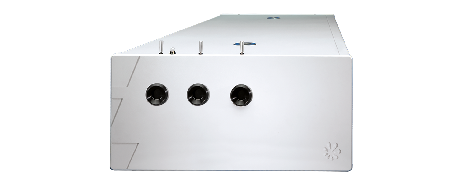
PhotoSonus T
High-energy tunable laser for photoacoustic imaging
The PhotoSonus T is the table-top version of the high-energy, tunable PhotoSonus M laser. It offers high output energies for imaging larger tissue volumes in the field of photoacoustic imaging.
Developed and manufactured by:
Features
- Software-controlled wavelength setting from 330 to 2600 nm
- Ultra-wide OPO signal tuning range from 660 to 1320 nm
- Up to 230 mJ in the 660 – 2600 nm range, 35 mJ in the 330 – 660 nm range
- Narrow line width over the entire tunable range
- Pulse duration of 3-5 ns
- Control via handheld device or PC
- Separate output port for 532 nm; optional output for 1064 nm
- Monitoring the OPO pump laser energy
- Fast wavelength change within the entire signal or idler range
Applications
- Photoacoustic imaging
- Flash photolysis
- Photobiology
- Remote Sensing
- Nonlinear spectroscopy
Applications
Hearing with light - seeing with sound
Laser-based photoacoustic imaging is an innovative method that combines light and sound to generate high-resolution images of biological tissue. Tissue is irradiated with short, usually spectrally tunable laser pulses. These lead to minimal local heating and thus generate ultrasound waves, which are measured and converted into images.
When light separates molecules
Photolysis describes the light-induced splitting of chemical compounds and plays a central role in photosynthesis through water splitting. Laser flash photolysis uses short, intense laser pulses (ns-fs) to generate short-lived intermediates such as radicals or excited states and to observe their temporally resolved absorption. Probe pulses record reaction processes. The use of OPOs significantly expands the possibilities of spectroscopic investigations.
Applications
Scientific publications
An Investigation of Signal Preprocessing for Photoacoustic Tomography
I. Huen, R. Zhang, R. Bi, X. Li, M. Moothanchery, and M. Olivo, Sensors 23 (1), 510 (2023). DOI: 10.3390/s23010510.
Photoacoustic tomography (PAT) is increasingly being used for high-resolution biological imaging at depth. Signal-to-noise ratios and resolution are the main factors that determine image quality. Various reconstruction algorithms have been proposed and applied to reduce noise and enhance resolution, but the efficacy of signal preprocessing methods which also affect image quality, are seldom discussed. We, therefore, compared common preprocessing techniques, namely bandpass filters, wavelet denoising, empirical mode decomposition, and singular value decomposition. Each was compared with and without accounting for sensor directivity. The denoising performance was evaluated with the contrast-to-noise ratio (CNR), and the resolution was calculated as the full width at half maximum (FWHM) in both the lateral and axial directions. In the phantom experiment, counting in directivity was found to significantly reduce noise, outperforming other methods. Irrespective of directivity, the best performing methods for denoising were bandpass, unfiltered, SVD, wavelet, and EMD, in that order. Only bandpass filtering consistently yielded improvements. Significant improvements in the lateral resolution were observed using directivity in two out of three acquisitions. This study investigated the advantages and disadvantages of different preprocessing methods and may help to determine better practices in PAT reconstruction.
Photoacoustic tomography with a model-based approach involving realistic detector properties
P. Warbal, and R. K. Saha, Results in Optics 13, 100528 (2023). DOI: 10.1016/j.rio.2023.100528.
A computational and experimental study is conducted to examine how directivity associated with a finite aperture sensor affects photoacoustic tomography (PAT) image reconstruction. Acoustic signals for the simulation work were computed using a discrete particle approach from three numerical phantoms including a vasculature. The theoretical framework and a Monte Carlo approach for construction of a tissue configuration are discussed in detail. While simulating forward data, the directivity of the sensor was taken into account. The image reconstruction was accomplished using system matrix based methods like l2 norm Tikhonov regularization, l1 norm regularization and total variation (TV) minimization. Accordingly, two different system matrices were constructed- (i) assuming transducer as a point detector (PD) and (ii) retaining properties of a finite detector with directivity (FDWD). Image reconstruction was also performed utilizing experimentally measured PA signals. Both the computational and experimental results demonstrate that blur-free PAT imaging can be achieved with the FDWD method. Additionally, TV minimization provides marginally better image reconstruction compared to the other schemes.
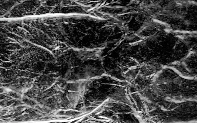
Fast photoacoustic imaging technology for deep structure information of finger
T. Meng, H. Li, and Y. Liu, in Ninth Symposium on Novel Photoelectronic Detection Technology and Applications, J. Chu, W. Liu, and H. Xu, eds. (SPIE, 2023), pp. 126176E. DOI: 10.1117/12.2666706.
In this paper, we exploited the fast-imaging technology for the deep structure of finger based on photoacoustic imaging, which adopted the self-designed 128-ring-array fast photoacoustic imaging system to acquire the latent inside information of finger. The home-made photoacoustic imaging system has the merits of fast imaging, high resolution and deep imaging depth. Specifically, our system could obtain a cross section scan of finger within 0.05 or 0.1s, achieve the resolution of approach 180 μm and image the latent inside information of finger as well as extend the imaging depth over 5 cm in chicken breast tissue at the laser density of 20 mJ/cm2 (≤ANSI safety limit). In this work, we obtained the finger anatomical information of skin tissue, blood vessel tissue, and the information of tendon tissue and phalanx tissue which is relatively difficult to obtain by means of photoacoustic imaging. Thus, we will be able to restore an overall internal structure of a finger including its external shape its internal tendon structure and its internal phalanx structure or containing its blood vessel structure. And that more information from different angles can make its identification more accurate. It is prospective that the deep structure of finger we get by our fast photoacoustic imaging technology will help to provide more possibilities for finger identification and lead to more credible technology for human about relevant information collection and resolution.