PhotoSonus M
High-energy, mobile, tunable laser source Photoacoustics
Due to increasing demand for high pulse energies for photoacoustic imaging to visualize larger tissue volumes, the PhotoSonus M was introduced – an advanced, high-energy, tunable laser source for photoacoustic imaging.
Developed and manufactured by:
Features
- High pulse energy up to 250 mJ
- Wide tuning range from 330 to 2300 nm
- Ultra-wide OPO signal tuning range from 660 to 1320 nm
- Pulse repetition rate of 10 Hz or 20 Hz
- Integrated pump laser, OPO and power supply in one mobile device
- Low maintenance costs
- Fiber connection with interlock
- Rapid wavelength change within the entire signal or idler range between two consecutive pulses
- Integrated energy meter (optional)
- Motorized attenuator (optional)
- Optional outputs for pump laser wavelengths 1064/532 nm
Highlights
- High pulse energy
- Mobile and low-maintenance
- Fiber connection
- Rapid change in wavelength
Application
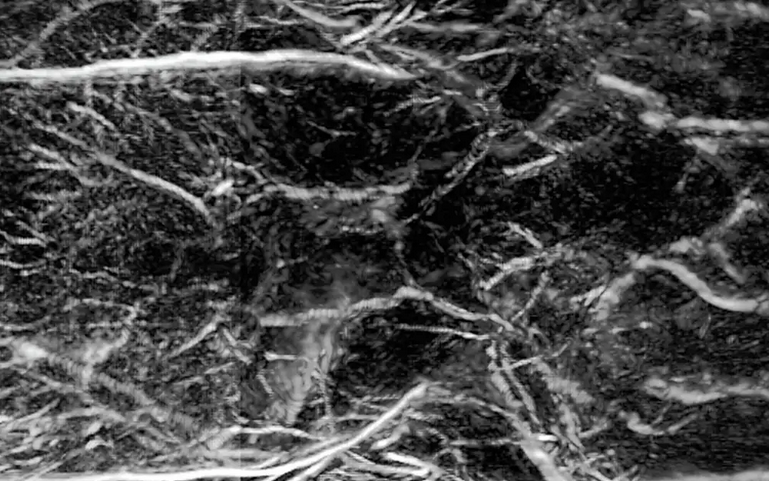
Hearing with light - seeing with sound
Laser-based photoacoustic imaging is an innovative method that combines light and sound to generate high-resolution images of biological tissue. Tissue is irradiated with short, usually spectrally tunable laser pulses. These lead to minimal local heating and thus generate ultrasonic waves, which are measured and converted into images.
Application
Scientific publications
Non-Invasive Photoacoustic Cerebrovascular Monitoring of Early-Stage Ischemic Strokes In Vivo
J. Kim, J. Y. Kweon, S. Choi, H. Jeon, M. Sung, R. Gao et al, Advanced Science 12 (4), 2409361 (2025). DOI: 10.1002/advs.202409361.
Early-stage stroke monitoring enables timely intervention that is crucial to minimizing neuronal damage and increasing the extent of recovery. By monitoring collateral circulation and neovascularization after ischemic stroke, the natural recovery process can be better understood, optimize further treatment strategies, and improve the prognosis. Photoacoustic computed tomography (PACT), a non-invasive imaging modality that captures multiparametric high-resolution images of vessel structures, is well suited for evaluating cerebrovascular structures and their function. Here 3D multiparametric transcranial PACT is implemented to monitor the early stage of a photothrombotic (PT)-stroke model in living rats. New vessels in the PT-induced region are successfully observed using PACT, and these observations are confirmed by histology. Then, using multiparametric PACT, it is found that the SO2 in the ischemic area decreases while the SO2 in newly formed vessels increases, and the SO2 in the PT region also recovers. These findings demonstrate PACT’s remarkable ability to image and monitor cerebrovascular morphologic and physiological changes. They highlight the usefulness of whole-brain PACT as a potentially powerful tool for early diagnosis and therapeutic decision-making in treating ischemic stroke.
Bimetallic Hyaluronate-Modified Au@Pt Nanoparticles for Noninvasive Photoacoustic Imaging and Photothermal Therapy of Skin Cancer
H. H. Han, S. Kim, J. Kim, W. Park, C. Kim, H. Kim et al, ACS Applied Materials & Interfaces 15 (9), 11609-11620 (2023). DOI: 10.1021/acsami.3c01858.
Although spherical gold (Au) nanoparticles have remarkable photothermal conversion efficiency and photostability, their weak absorption in the near-infrared (NIR) region and poor penetration into deep tissues have limited further applications to NIR light-mediated photoacoustic (PA) imaging and noninvasive photothermal cancer therapy. Here, we developed bimetallic hyaluronate-modified Au-platinum (HA-Au@Pt) nanoparticles for noninvasive cancer theranostics by NIR light-mediated PA imaging and photothermal therapy (PTT). The growth of Pt nanodots on the surface of spherical Au nanoparticles enhanced the absorbance in the NIR region and broadened the absorption bandwidth of HA-Au@Pt nanoparticles by the surface plasmon resonance (SPR) coupling effect. In addition, HA facilitated the transdermal delivery of HA-Au@Pt nanoparticles through the skin barrier and enabled clear tumor-targeted PA imaging. Compared to conventional PTT via injection, HA-Au@Pt nanoparticles were noninvasively delivered into deep tumor tissues and completely ablated the targeted tumor tissues by NIR light irradiation. Taken together, we could confirm the feasibility of HA-Au@Pt nanoparticles as a NIR light-mediated biophotonic agent for noninvasive skin cancer theranostics.
Characterizing a photoacoustic and fluorescence imaging platform for preclinical murine longitudinal studies
W. R. Thompson, H. F. Brecht, V. Ivanov, A. M. Yu, D. S. Dumani, D. J. Lawrence et al, Journal of Biomedical Optics 28 (3), 036001 (2023). DOI: 10.1117/1.JBO.28.3.036001.
Significance. To effectively study preclinical animal models, medical imaging technology must be developed with a high enough resolution and sensitivity to perform anatomical, functional, and molecular assessments. Photoacoustic (PA) tomography provides high resolution and specificity, and fluorescence (FL) molecular tomography provides high sensitivity; the combination of these imaging modes will enable a wide range of research applications to be studied in small animals.
Aim. We introduce and characterize a dual-modality PA and FL imaging platform using in vivo and phantom experiments.
Approach. The imaging platform’s detection limits were characterized through phantom studies that determined the PA spatial resolution, PA sensitivity, optical spatial resolution, and FL sensitivity.
Results. The system characterization yielded a PA spatial resolution of 173 ± 17 μm in the transverse plane and 640 ± 120 μm in the longitudinal axis, a PA sensitivity detection limit not less than that of a sample with absorption coefficient μa = 0.258 cm – 1, an optical spatial resolution of 70 μm in the vertical axis and 112 μm in the horizontal axis, and a FL sensitivity detection limit not <0.9 μM concentration of IR-800. The scanned animals displayed in three-dimensional renders showed high-resolution anatomical detail of organs.
Conclusions. The combined PA and FL imaging system has been characterized and has demonstrated its ability to image mice in vivo, proving its suitability for biomedical imaging research applications.